 Using ultrasound to look inside the womb at the growing baby is almost as exciting as the pregnancy itself. You can find out if it's a boy or a girl, as well as a host of other facts. You can even see if the baby is a thumbsucker, calm or energetic. Most importantly, an ultrasound scan can reassure you that the baby is healthy and growing properly.
In addition to radio and television, which use a spectrum of frequencies that we can hear, there are devices that use frequencies beyond our hearing range. These frequencies are called ultrasound, radar or sonar. Ultrasound, or medical sonography, was developed by a doctor in the 1960s and was inspired by a dental instrument that used ultrasound to clean teeth.
Medical sonography works like other ultrasound techniques. It generates a sound wave that bounces off objects and registers the reflected sound. As a diagnostic tool, ultrasound is non-invasive, gives clear results and poses no risk to the patient when used by a trained practitioner. The technology, unlike MRI or X-ray machines, is fairly easy to use and relatively inexpensive.

The basis for medical ultrasound examinations begins with a study by the Curie brothers on the properties of crystals in the nineteenth century. Their discovery that the compression of certain crystals produces an electrical discharge led to the study of sound waves that enabled sonar in World War I, radar in World War II, and the dental plaque blaster. Ultrasound begins with a piezoelectric transducer, an instrument that compresses various types of quartz or ceramic between metal plates until it emits high-frequency sound waves. Most medical ultrasound transducers use ceramic that emits a frequency wave between 2 and 18 megahertz. A technician directs the sound with a wand-like device that emits the sound and receives the echo as the sound bounces off internal organs, tissues and bones. A warming gel on the patient's skin prevents false echoes from the skin itself.
The type and quality of the return sound defines the distance from the transducer and its shape and density. The vibration of the transducer plates generates electrical current, which is translated into a digital signal in the scanner. Software then translates the signals into an image and data. As the sound waves are emitted from a source, the resulting images are cone-shaped.
Medical sonography is most commonly associated with the development of the foetus in the womb due to its ease of use and safety. Sonograms can detect congenital physical problems as well as potential multiple births and incorrect foetal position. Sonograms are commonly used in gynaecological and oncological examinations and can be used in places where visual examinations are problematic.
Modern ultrasound technology has developed applications of Doppler theory that show motion, velocity and sound.
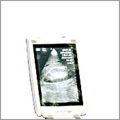
General Electric' has just developed a mobile ultrasound device that is little bigger than a foldable mobile phone. This sensational device is expected to greatly improve diagnostic capabilities in the near future as doctors can literally carry it in their pocket. Referrals to specialists should be reduced as this portable ultrasound device will be more accessible to doctors. It is hoped that this is the first step towards an easy-to-use ultrasound machine for home use.